Ensure consistency and reproducibility of results through our in vitro cell culture solutions

Cell Culture

お問い合わせ

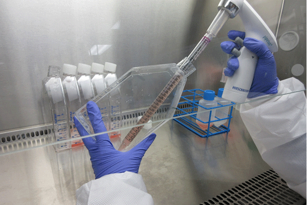
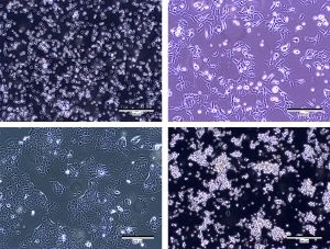
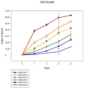
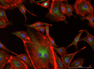
Whether you need to grow immortalized tumor cells, primary cells, immune cells, neurospheres or spheroids for your study, our team can grow them. Our cell culture and in vitro experts have diverse overlapping skills to grow cells in vitro and perform numerous types of downstream treatments and assays with the cells. Our experience using multiple types of incubators, vessels, and systems to grow cells and keep them healthy means we are the CRO of choice for cell culture. Choose from one of the 400 cell lines in our repository – which includes over 100 Luciferase-enabled for easy monitoring – or 3D spheroids to better predict clinical outcomes and consider us your choice for your next preclinical oncology project.


  • 3D cell culture

    Our mission is to reduce the failure rate of candidate anticancer drugs in clinical trials by providing our clients with access to advanced tumor-specific 3D in vitro models. Predictive Oncology® 3D in vitro models provide us with a new tool that produces more reliable outcomes for early evaluation of responses to oncology and immuno-oncology therapies. This comprehensive 3D culture platform integrates organ-specific cellular and extracellular elements to maintain the critical interactions between a tumor and its surroundings allowing you to eliminate ineffective compounds early in the drug development process and move forward with promising candidates.

  • Large-scale culture

    Scientist working on a large scale analysis. Scientist is holding a container and a scientific mechanism

    Labcorp cell culture analysts have experience with over 500 cell lines, including models for over 75 histotypes, covering acute monocytic leukemia (AML) to vulva leiomyosarcoma. We have an experienced team of analysts who are familiar with various mechanisms for cell culture scale up including culture in plates, flasks, roller bottles, spinner flasks, and multi-layered flasks. Importantly, we have capacity to process thousands of flasks per week to support large- and small-scale animal work including support for over 40 models on our FastTrack program, which includes initiation of cell culture within 2 weeks of study signing.


  • 細胞形態

    Labcorp can monitor the cell morphology of your cells in culture using an Olympus CKX53F inverted fluorescence microscope with phase contrast and a 1.4MP CCD camera. 治療の前後や治療中に、長期にわたって、あるいは移植前の特定のタイミングでも画像をチェックし、細胞が正常に成長していることを確認しましょう。

  • 細胞増殖 / 腫瘍形成

    We o   can monitor the growth of your cells over time and compare different cell variants, cells grown in different media, or cells grown with different treatments. We have two Luna CellDrop Automated Cell Counters that can accurately distinguish the live from the dead cells using trypan blue or AO/PI and count each population. Our cell culture team has a vast amount of experience culturing multiple types of cells including immortalized tumor cells, primary cells, immune cells, neurospheres, and spheroidstumor cells, primary cells, and neurospheres. The team also has experience performing colony formation assays to detect the tumorigenic potential of transformed cells and the tumor suppressive effects of proteins on transformed cells in vitro. Our flow cytometry team has experience monitoring the proliferation and viability of stimulated T- cells or T- cells in mixed lymphocyte reactions and co-cultures using CFSE staining or other cell tracking reagents.

  • High content image analysis

    Labcorp has the ability to stain cultured 2D or 3D cells and acquire fluorescent images using the Cytation 3 Imaging Plate Reader. 画像が撮影されると、特殊ソフトウェアを使って核、生細胞と死細胞、染色細胞(陽性 / 陰性別)の数を計上し、サイズ、真円度、面積、周囲、各色の強度を測定します。

  • 画像化可能な細胞株の作成

    ホタル(P. pyralis、フォティヌス・ピラリス)のルシフェラーゼは、D-ルシフェリンがあると生物発光を起こす酵素です。ルシフェラーゼ 2 (luc2) 遺伝子の DNA を、レンチウイルス形質導入システムを用いて、ピューロマイシン耐性遺伝子とともに、細胞のゲノム中に組み込みます。細胞を培養している間にピューロマイシンを培地に加え、ルシフェラーゼ遺伝子が組み込まれた細胞のみを選択できるようにします。 Once the luc-enabled cells are injected into a mouse or rat and D-luciferin is administered, the light produced can be detected with Labcorp's IVIS® Spectrum in vivo imaging system. The in vitro services team has experience performing every step of the process from stably virally transducing cells, to selecting the transduced cells, to measuring the light output of the cells.


  • Cytotoxicity/proliferation assay


    細胞毒性 / 増殖アッセイは、化合物ライブラリの細胞毒性スクリーニングで広く使用されています。 If you are interested in developing a therapeutic agent that targets rapidly dividing cancer cells, we can test the potency of your cytotoxic compounds.当社では、医薬品への投資に先立って、初回のハイスループットスクリーニングで、望ましくない細胞毒性効果を持つ可能性の高い「ヒット」を選別することも可能です。 Labcorp can measure the toxicity of your compound in your choice of over nearly 400 cell lines that we have availablestock in our repository. We provide graphs and IC50 values for each cell line and compound.

お問い合わせはこちら

お問い合わせ